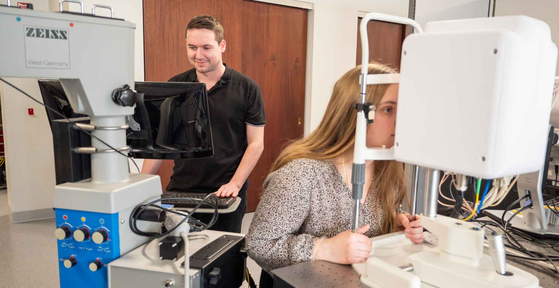
Intraoperative Fluorescence Nerve Imager
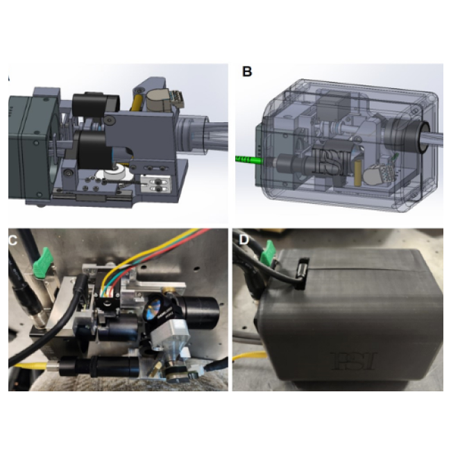
PSI has developed an intraoperative fluorescence imager that is capable of identifying nerve tissue in the surgical bed. The PSI fluorescence imager uses an innovative tempo-spatial modulation technique to reject the ambient light background, eliminating the need for turning off the surgical lighting during fluorescence image acquisition. This significantly reduces the interruption to the normal surgical workflow, lowering the barrier for clinical adoption.
High-contrast nerve highlighting images with a high background rejection ratio help surgeons avoid iatrogenic nerve injury during surgery.
